Assessment of the P-wave is essential for diagnosing various cardiac arrhythmias, atrial abnormalities, and conduction disturbances. It helps clinicians evaluate atrial function and detect conditions such as atrial fibrillation, atrial flutter, atrial enlargement, and atrial ischemia.

Duration
<110 ms
Amplitude
< 2,5 mm in limb leads
< 1,5 mm in precordial leads
Measurement
lead II for P wave duration
V1 is used to calculate the negative P wave terminal force
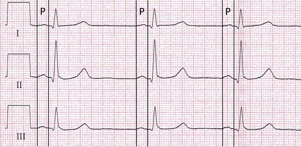
Normal P-wave. Despite often being simplified as smooth and rounded, the P wave carries a significant amount of information beyond this basic description. The variations in morphology, including subtle beta-to-beta changes, can provide valuable insights into cardiac conduction and atrial function. The P wave is frequently underestimated in its significance within the ECG interpretation process. Limb leads I – III, 50 mm/s.
Definition
In the presence of sinus rhythm, the P-wave is indeed the first deflection observed on an electrocardiogram (ECG) tracing. It represents the depolarization (activation) of the atria as the electrical impulse spreads from the sinoatrial (SA) node throughout the atrial myocardium.
Duration
The normal duration of the P-wave is typically between 80 to 110 milliseconds in duration. P-waves that exceed this duration may indicate abnormal atrial depolarization. The terminal negative deflection of a biphasic P wave in lead V1 is referred to as the P terminal force V1 (PTFV1). PTFV1 is calculated as the product of its duration component (PTDV1) and its absolute amplitude component (PTAV1). An absolute PTFV1 value ≥4 mV·ms is considered abnormal.2,3 PTFV1 is generally perceived as a marker of left atrial pathology, and abnormal PTFV1 has been associated with incident atrial fibrillation4, 5, 6 and stroke.
Amplitude:
The amplitude of the P-wave refers to the height of the waveform measured from the baseline (isoelectric line) to the peak of the deflection. In standard limb leads, the normal amplitude of the P-wave is generally less than 2.5 mm, while in precordial leads, it is less than 1.5 mm.
Shape
In the limb leads, the shape of the P-wave is typically smooth and rounded, resembling a gentle curve. However, variations in shape can occur depending on individual differences, lead placement, and underlying cardiac conditions.
Measurement:
Lead II is often used to assess the P-wave morphology during sinus rhythm because it provides a good view of atrial depolarization. In a normal ECG, the P-wave in lead II is typically upright (positive) and precedes the QRS complex.
The terminal negative deflection of a biphasic P wave in lead V1 is referred to as the P terminal force V1. It is calculated as the product of its duration component and its absolute amplitude component. An absolute value ≥4 mV·ms is considered abnormal. P terminal force V1 is generally perceived as a marker of left atrial pathology, and abnormal value has been associated with incident atrial fibrillation and stroke.
Clinical Significance:
Assessment of the P-wave is essential for diagnosing various cardiac arrhythmias, atrial abnormalities, and conduction disturbances. It helps clinicians evaluate atrial function and detect conditions such as atrial fibrillation, atrial flutter, atrial enlargement, and atrial ischemia.
References
- Davies HJ, Hammour G, Zylinski M, et al. The Deep-Match Framework: R-Peak Detection in Ear-ECG. IEEE Trans Biomed Eng. 2024 Jan 29;PP.
- Jafari Afshar E, Gholami N, Samimisedeh P, et al. Utility of electrocardiogram to predict the occurrence of the no-reflow phenomenon in patients undergoing primary percutaneous coronary intervention (PPCI): a systematic review and meta-analysis. Front Cardiovasc Med. 2024;10:1295964
- Tereshchenko LG. P-terminal force in lead V1: May the force be with you. Heart Rhythm. 2023 Mar;20(3):363-364.
Related Pages
Atrial tachycardia | atrial fibrillation | atrial flutter | atrial fibrillation | atrial flutter | atrial fibrillation | atrial flutter | atrial fibrillation | atrial flutter | atrial fibrillation | atrial flutter | atrial fibrillation | atrial flutter
